Echocardiography
Detailed, real-time imaging of the heart’s structure and function
Echocardiography is a safe, non-invasive ultrasound technique that provides detailed images of the heart’s chambers, valves, walls, and surrounding structures. It plays a vital role in diagnosing a wide range of cardiac conditions and in monitoring how the heart responds to treatment over time.
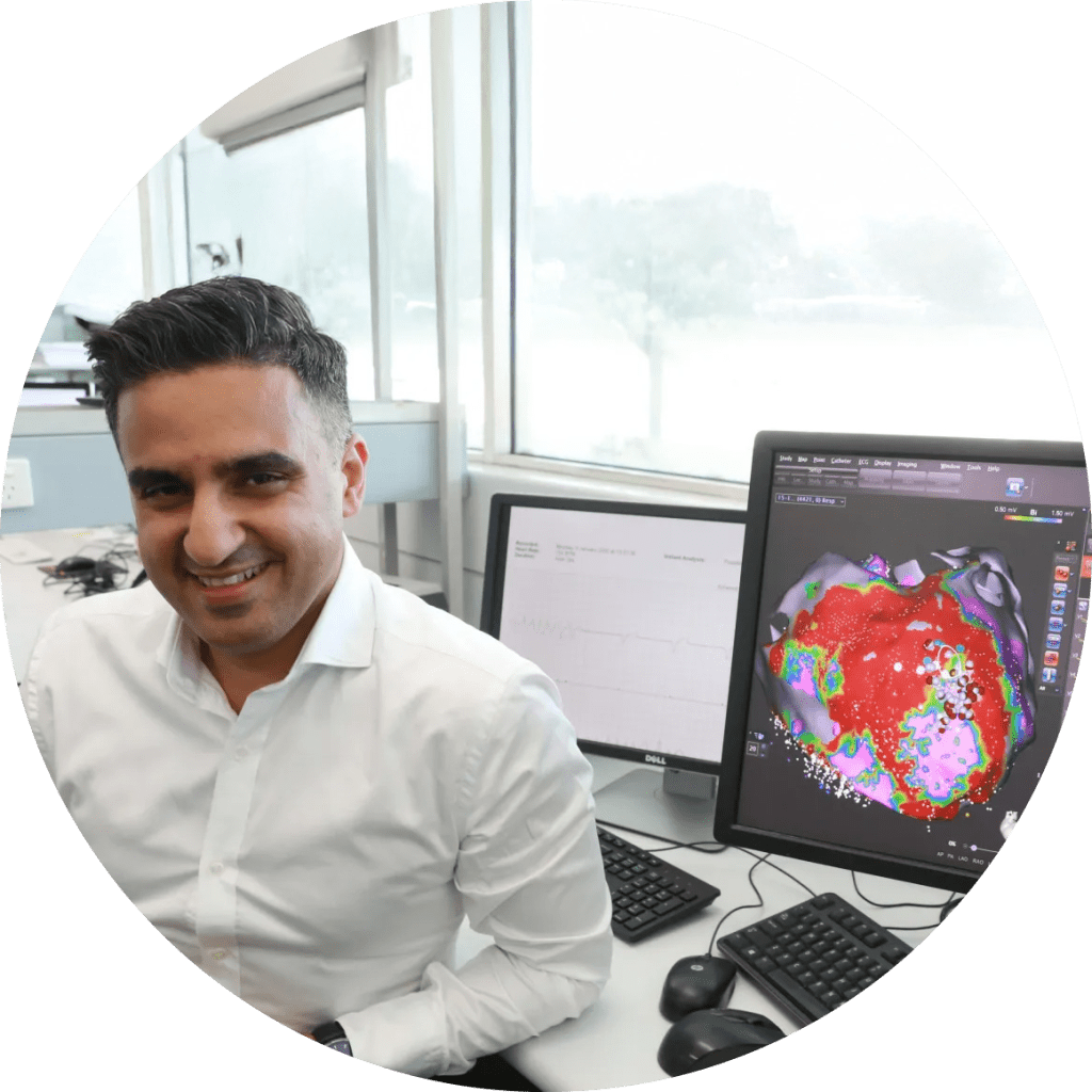
At his Sydney-based practice, A/Prof Saurabh Kumar offers expert echocardiographic evaluation as part of comprehensive cardiology care. With over 15+ years of experience and a subspecialty in interventional electrophysiology, A/Prof Kumar uses echocardiography not only to assess structural heart disease but also to guide rhythm-related investigations and long-term cardiac management.
When Is Echocardiography Recommended?
Heart failure
Echocardiography helps determine whether heart failure is due to impaired contraction (reduced ejection fraction) or impaired relaxation (preserved ejection fraction), and informs treatment decisions.
Valvular heart disease
Congenital heart abnormalities
Including structural defects present from birth, such as septal defects or abnormal connections between heart chambers or vessels.
Pericardial effusion
Accumulation of fluid around the heart can be detected and monitored with echocardiography, helping to prevent complications such as cardiac tamponade.
Cardiomyopathy
Includes hypertrophic (HCM), dilated (DCM), and restrictive (RCM) forms of heart muscle disease. Echo provides essential information on chamber size, wall thickness, and overall function.
Atrial and ventricular septal defects
These are openings between the heart’s chambers that may be congenital or acquired. Echo, especially with contrast or transoesophageal imaging, helps define the size and clinical significance of these defects.
Echocardiography is often the first test requested when a heart murmur is heard, when a patient presents with shortness of breath, or when an abnormal ECG or chest X-ray raises concern about structural heart disease.

Types of Echocardiographic Services Offered
A/Prof Kumar offers a full suite of echocardiographic services, using both transthoracic and transoesophageal techniques, depending on the clinical indication.
Transthoracic Echocardiogram (TTE)
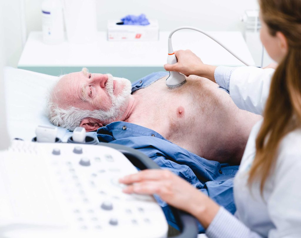
The standard and most commonly used form of echocardiography. It involves placing an ultrasound probe on the chest wall to generate real-time images of the heart’s structures. TTE is painless, radiation-free, and typically takes 30 to 45 minutes.
It is useful for:
- Assessing heart chamber size and function
- Evaluating valve anatomy and flow patterns
- Measuring wall thickness and motion
- Detecting pericardial fluid
- Estimating pressures inside the heart and pulmonary arteries
Transoesophageal Echocardiogram (TOE)
In some cases, clearer images are needed—particularly of structures at the back of the heart such as the left atrial appendage or atrial septum. TOE involves passing a specialised probe down the oesophagus under sedation. This provides closer and more detailed views, especially useful in:
- Detecting clots before cardioversion for atrial fibrillation
- Evaluating prosthetic heart valves
- Diagnosing infective endocarditis
- Identifying atrial septal defects or patent foramen ovale
TOE is performed in coordination with hospital imaging teams and under safe, monitored conditions.
Bubble Contrast Studies
Serial Imaging for Chronic Cardiac Conditions
Ejection Fraction (EF) Assessment
Ejection fraction is a key measurement that reflects how well the heart is pumping. It is critical in diagnosing and managing heart failure, monitoring recovery post-heart attack, and determining eligibility for certain therapies such as implantable defibrillators or cardiac resynchronisation therapy.
Assessment of Chamber Sizes and Wall Motion
The Echocardiography Experience
Echocardiography is a simple, well-tolerated procedure. For transthoracic echo, patients lie on a bed while a technician or cardiologist uses a handheld probe with ultrasound gel to scan the heart from various angles. The test is safe, non-invasive, and does not require fasting or recovery time.
In contrast, transoesophageal echo involves mild sedation, and patients are typically advised to fast beforehand. A/Prof Kumar will explain the purpose and process clearly in advance and answer any questions you may have.
All echocardiographic images are analysed in detail by A/Prof Kumar himself, ensuring that the results are interpreted within the full context of your clinical history and cardiac care plan.


Take Control of Your Heart Health Today.
A/Prof Saurabh Kumar brings over 15+ years of clinical expertise to the care of patients with heart rhythm disorders and general cardiac conditions. He is widely regarded within the Australian cardiology community and internationally for his depth of knowledge, collaborative style, and commitment to patient-centred care.
He holds dual roles as a Staff Specialist Cardiologist and Cardiac Electrophysiologist at Westmead Hospital and Clinical Associate Professor of Medicine at the University of Sydney. He currently serves as the Program Director for Ventricular Arrhythmias and Sudden Cardiac Death at Westmead Hospital and is the Translational Electrophysiology Lead at the Westmead Applied Research Centre, University of Sydney.